Article

Isolated and Combined Effect of Age and Gender on Neutrophil–Lymphocyte Ratio in the Hyperglycemic Saudi Population

1
Chair of Medical and Molecular Genetics Research, Department of Clinical Laboratory Sciences, College of Applied Medical Sciences, King Saud University, Riyadh 12372, Saudi Arabia
2
Department of Clinical Laboratory Sciences, College of Applied Medical Sciences, King Saud University, Riyadh 12372, Saudi Arabia
3
Department of Clinical Laboratory Sciences, College of Applied Medical Sciences, King Khalid University, Abha 61421, Saudi Arabia
4
Department of Clinical Biochemistry, Faculty of Medicine, King Abdulaziz University, Jeddah 21589, Saudi Arabia
5
Department of Clinical Pathology, Al-Borg Medical Laboratories, Jeddah 23437, Saudi Arabia
6
Ministry of Health, Jeddah 21176, Saudi Arabia
*
Author to whom correspondence should be addressed.
Medicina 2022, 58(8), 1040; https://doi.org/10.3390/medicina58081040
Submission received: 15 June 2022 / Revised: 14 July 2022 / Accepted: 25 July 2022 / Published: 3 August 2022
(This article belongs to the Section Endocrinology)

Abstract

:
Inflammation is pivotal to the pathogenesis of diabetes mellitus (DM), but pathological alterations of the neutrophil–lymphocyte ratio (NLR), an emerging inflammatory index in DM management, remains understudied. The aim of this study is to examine the relationship between NLR and glycemic control in the Saudi population. Gender, age, WBC count, and fasting blood glucose (FBG) were obtained from Al-Borg Medical Laboratories for 14,205 subjects. Means, prevalence, risk measures, and the diagnostic accuracy of elevated NLR and hyperglycemia (HG) were evaluated. Subjects with elevated NLR (>3) had significantly higher FBG (105.10 ± 0.33 vs. 114.0 ± 2.81) and NLR was significantly elevated in impaired fasting glycemia (IFG; 1.21 ± 0.01 vs. 1.25 ± 0.01) and HG (1.21 ± 0.01 vs. 1.39 ± 0.02). Elevations of NLR in HG but not in IFG persisted across all age groups except young males and elderly females. The prevalence of elevated NLR in hyperglycemic subjects was 4.12% compared to 2.16% in subjects with normal FBG. HG was more prevalent in subjects with elevated NLR (17.33% vs. 12.46%) who had a relative risk (RR) of 1.68 (95% CI = 1.38–2.06, p < 0.0001) and an odds ratio (OR) of 1.94 (95% CI = 1.48–2.56, p < 0.0001) to be hyperglycemic. Nevertheless, NLR failed to discriminate individuals with normal FBG from those with HG based on ROC curve analysis. Pathological fluctuations in NLR may serve as supportive evidence in DM management.

1. Introduction

Diabetes mellitus (DM) is a metabolic disease whose pathognomonic manifestation is persistent hyperglycemia (HG) defined by the American Diabetes Association (ADA) as fasting blood glucose (FBG) ≥126 mg/dL [1]. Impaired fasting glycemia (IFG), defined as FBG of 100–125 mg/dL, precedes, and may progress to, overt HG if left untreated [2]. Risk factors for DM include genetic predisposition [3], sedentary lifestyle [4], dietary habits [5], and tobacco smoking [6]. Notably, the interplay between insulin resistance and inflammation contributes to the cytokine and adipokine surge characteristic of DM pathogenesis [7]. Consequently, uncontrolled DM is often complicated by inflammatory conditions, including vasculopathy, retinopathy, nephropathy, and neuropathy [8].
Impaired insulin secretion in type 1 DM (T1DM) to some extent is due to destruction of β-cells by TNF-α, IFN-γ, and IL-1β [9]. In addition to releasing proinflammatory cytokines, macrophages also participate in islet inflammation by creating an oxidative milieu through which cellular injury ensues [10]. Moreover, β-cell death may be instigated by inflammatory infiltrate of T lymphocytes as studied in diabetic mice and humans. Other immune cells; namely, natural killer (NK) cells, NKT cells, and dendritic cells, may similarly modulate T1DM development and progression [11]. In type 2 DM (T2DM), chemotactic white blood cells (WBCs) contribute to vascular injury precipitated by oxidative stress and cytokine release [12]. Although the WBC count has a predictive value for T2DM complications [13], the neutrophil–lymphocyte ratio (NLR), an emerging biomarker, has significant prognostic and stratifying superiority over traditional WBC count in a wide spectrum of inflammatory conditions [14].
NLR represents the absolute counts of circulating neutrophils and lymphocytes, and increased NLR may thus be caused by either elevated neutrophils and/or diminished lymphocytes. As such, NLR serves as a marker of systemic inflammation and disturbed cell-mediated immunity. In theory, physiological stress and pathological conditions in which inflammation or impaired immunity are central to the underlying pathophysiology would result in disrupted NLR. Indeed, investigators have explored the value of NLR in the differential diagnosis of conditions with a similar clinical picture [15,16] and in prognostication of a myriad of diseases [17,18]. Previous reports have found associations between increased NLR and the severity of metabolic syndrome, appendicitis, sepsis, COVID-19, and inflammatory bowel disease, as well as outcome and mortality in tuberculosis, cardiovascular disease, and cancer [19,20,21,22]. In the case of DM, elevated circulating neutrophils may be secondary to inordinate bone marrow production, or a return of resident cells in tissues to the blood [23]. Additionally, interleukins have been shown to cause neutrophilia and lymphopenia during inflammation [24] resulting in elevated NLR.
Because neutrophils utilize glucose as the major source of energy, persistent HG and advanced glycation end (AGE) products drive neutrophil dysfunction in DM. This manifests as impaired defense mechanisms, such as phagocytosis and oxidative burst, which increases susceptibility to infection, as well as augmented NETosis and cytokine release, further exacerbating inflammatory damage and islet cell death in DM [25]. Moreover, HG gives rise to hyperosmotic stress which in turn leads to intracellular Ca2+ accumulation, loss of cellular volume, and aberrant neutrophil function [26]. The diabetic microenvironment is also rich in AGE which are potent inducers of reactive oxygen species and upregulate pyroptosis-related mediators in neutrophils [27,28]. Other contributors to neutrophil dysfunction include lipids, infectious and chronic conditions, and aging [25]. Along those lines, a positive association between NLR and glycated hemoglobin A1c (HbA1c) [29] and between NLR and DM complications, including albuminuria and retinopathy, have been established [30,31].
Several lines of evidence point to a promising role of NLR in DM diagnosis and management; nonetheless, population-based studies on the patterns of NLR in HG are severely lacking. Furthermore, the effect of age and gender on the association between NLR and FBG remains understudied, especially in the Saudi population. Thus, the aim of this study is to assess the relationship between NLR and FBG by examining prevalence rates, association estimates, and the diagnostic accuracy of NLR for HG in gender- and age-adjusted analyses.

2. Materials and Methods

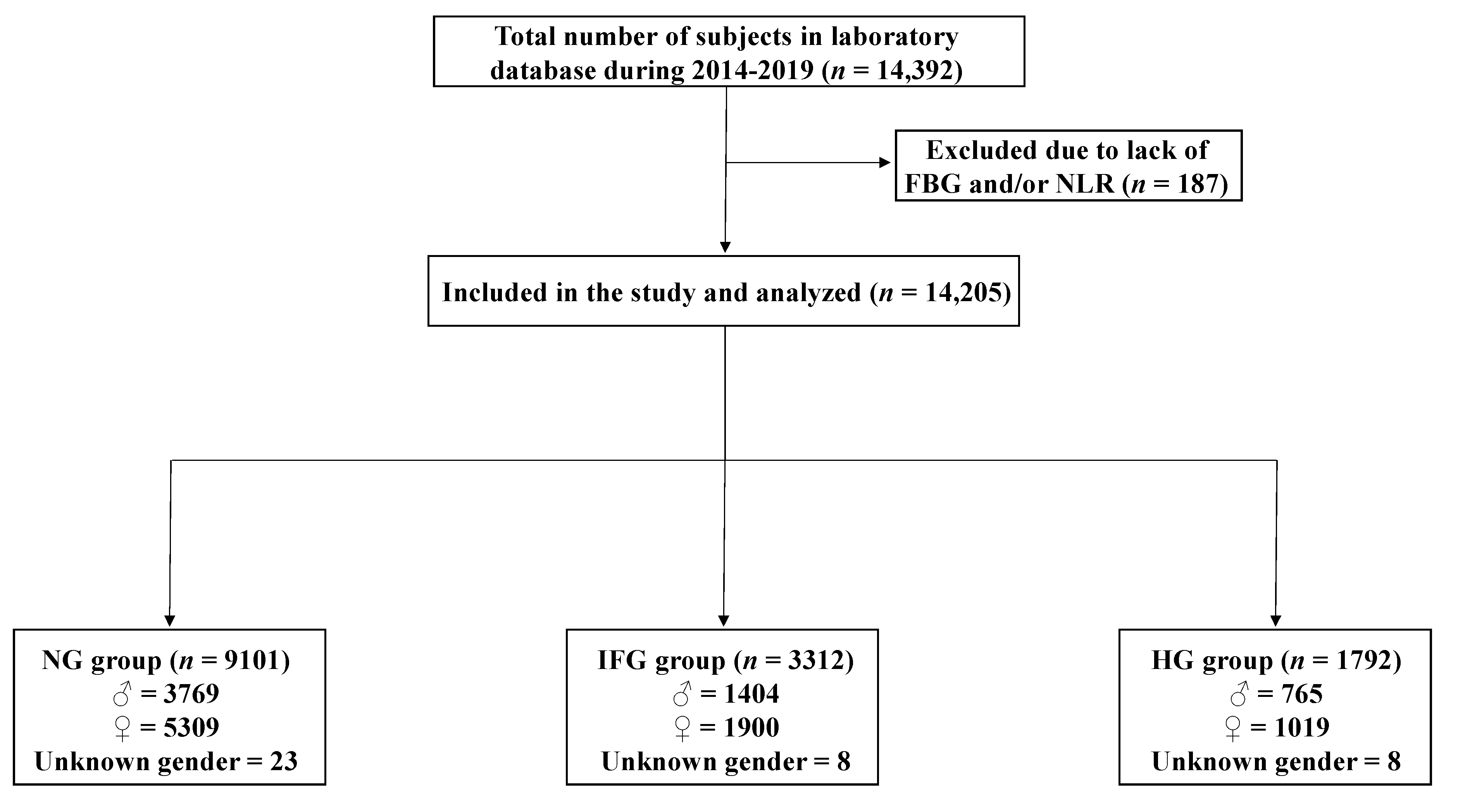
2.1. Study Design, Population, and Data Collection

Following approval of the Biomedical Ethics Unit of Al-Borg Medical Laboratories, Jeddah, Saudi Arabia, gender, age, and laboratory data for 14,205 subjects, collected during 2014–2019, were retrieved from Al-Borg Medical Laboratories database and retrospectively analyzed (Figure 1). Subjects were either walk-ins or referred to the laboratory by a physician. Those with missing information required for a particular analysis were excluded. Subjects were stratified based on gender and age as shown in Table 1.
Blood samples were collected once for each subject in vacutainer tubes coated with sodium fluoride and EDTA for FBG measurement and WBC count, respectively. Subjects were stratified based on FBG cutoffs published by the ADA [1]. Normal glucose (NG) was defined by values <100 mg/dL, IFG by 100–125 mg/dL, and HG by ≥126 mg/dL. NLR ranging from 1 to 3 was considered normal [32].

2.2. Statistical Analysis

Data are shown as means ±95% CI (in figures) or SEM (in text) as indicated. Means were compared by either unpaired, two-tailed student’s t-test for two groups or one-way ANOVA followed by Tukey’s post-hoc test for three or more groups. Association between FBG and NLR was tested by Pearson correlation and simple linear regression, and risk assessment measures; namely, the relative risk (RR), odds ratio (OR), and absolute risk reduction (ARR), were calculated as permitted by the natural sampling design of the study. All analyses were carried out using GraphPad Prism v9.2.0 (GraphPad Software, Inc., San Diego, CA, USA), and significance was defined by a p value of <0.05.

3. Results

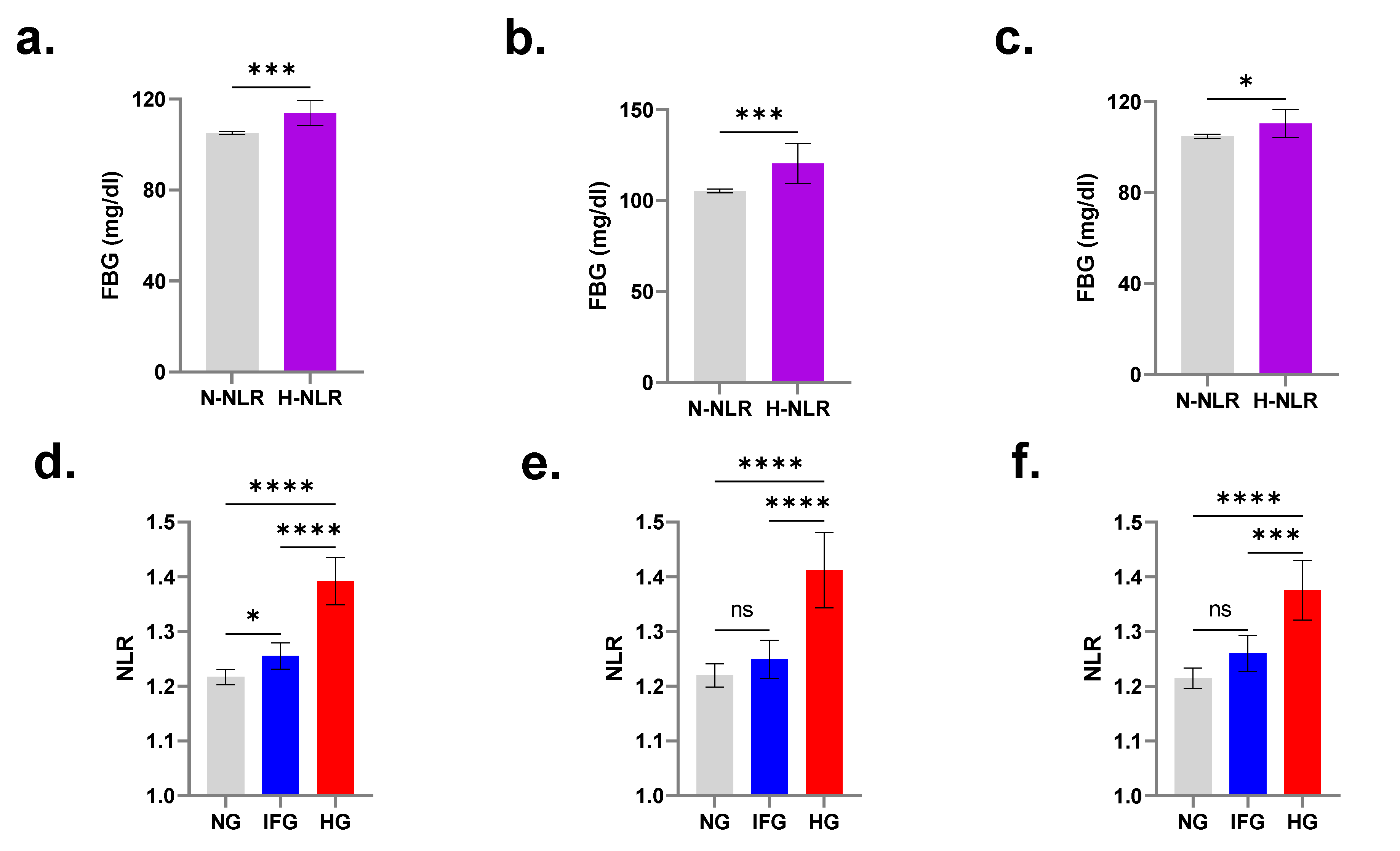
3.1. FBG Is Significantly Increased in Individuals with Elevated NLR

The total number of participants in our population was 14,205, of whom NG were 9101, IFG were 3312, and HG were 1792. In order to assess FBG levels in light of NLR, subjects of both genders and across all age groups were defined as either of normal NLR (N-NLR) or high NLR (H-NLR). As shown in Figure 1, FBG was consistently significantly increased in the H-NLR group in both genders (Figure 2a; 105.10 ± 0.33 mg/dL vs. 114.0 ± 2.81 mg/dL), in males (Figure 2b; 105.4 ± 0.5 mg/dL vs. 120.4 ± 5.5 mg/dL), and in females (Figure 2c; 104.8 ± 0.43 mg/dL vs. 110.4 ± 3.12 mg/dL).

3.2. Subjects with IFG and HG Have Significantly Elevated NLR

Figure 2d shows that those with normal FBG of both genders had an NLR of 1.21 ± 0.01 which significantly increased to 1.25 ± 0.01 in IFG and to 1.39 ± 0.02 in HG. When males and females were considered separately, NLR failed to distinguish between participants with normal FBG and those with IFG. However, it remained significantly elevated in hyperglycemic males (1.22 ± 0.01 vs. 1.41 ± 0.04) and females (1.21 ± 0.01 vs. 1.37 ± 0.03) as shown in Figure 2e,f, respectively.

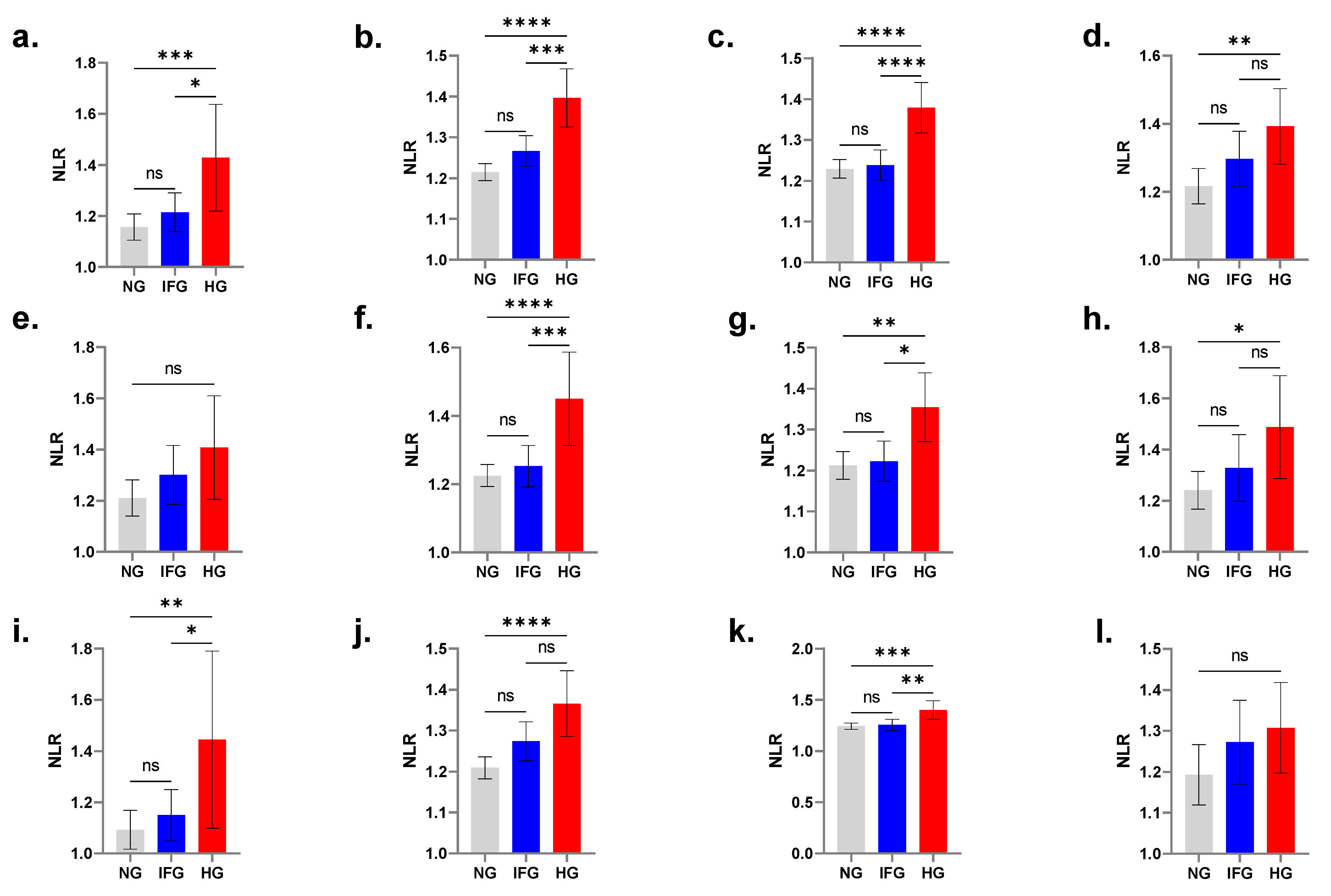
3.3. Age-Controlled Comparisons Displace NLR Significance in IFG

In both genders, NLR was significantly elevated in hyperglycemic young subjects (Figure 3a; 1.15 ± 0.03 vs. 1.42 ± 0.11), young adults (Figure 3b; 1.21 ± 0.01 vs. 1.39 ± 0.04), adults (Figure 3c; 1.22 ± 0.01 vs. 1.37 ± 0.04), and elderlies (Figure 3d; 1.21 ± 0.03 vs. 1.39 ± 0.06) compared to their normal counterparts.

3.4. NLR Is Not Elevated in Hyperglycemic Young Males

Combined age- and gender-wise comparisons revealed that NLR failed to discriminate young males with and without HG as shown if Figure 3e (1.21 ± 0.04 vs. 1.41 ± 0.1). Significantly increased NLR was observed in males with HG across all other age groups including young adults (Figure 3f; 1.22 ± 0.02 vs. 1.45 ± 0.07), adults (Figure 3g; 1.21 ± 0.02 vs. 1.35 ± 0.04), and elderlies (Figure 3h; 1.24 ± 0.04 vs. 1.48 ± 0.10).

3.5. NLR Is Not Elevated in Hyperglycemic Elderly Females

Significantly increased NLR was detected in females with HG in the young (Figure 3i; 1.10 ± 0.04 vs. 1.44 ± 0.17), young adult (Figure 3j; 1.21 ± 0.01 vs. 1.36 ± 0.04), and adult (Figure 3k; 1.24 ± 0.02 vs. 1.40 ± 0.05) age groups. However, NLR failed to discriminate elderly females with and without HG, as depicted in Figure 3l (1.19 ± 0.04 vs. 1.31 ± 0.1).

3.6. Elevated NLR Is More Prevalent in HG Subjects

The overall prevalence of elevated NLR among all subjects was 3.01%. Table 2 demonstrates that this rate decreased to 2.16% in subjects with normal FBG and increased to 4.71% in IFG and to 4.12% in hyperglycemic participants. A more profound pattern of increase was observed when either gender was considered alone. Likewise, HG was more prevalent in H-NLR compared to N-NLR subjects (17.33% vs. 12.46%) which, again, was consistent in either gender.

3.7. Elevated NLR Carries a Greater Risk for HG

As revealed in Table 3, H-NLR males were 2.60 times more likely to have HG and had 3.77 times the chance of having HG compared to males with N-NLR. Similarly, H-NLR females were 2.63 times more likely to have HG and had 3.73 times the chance of having HG compared to females with N-NLR. Additionally, ARR was −4.87% in both genders, −16.58% in males, and −15.59% in females.

3.8. Correlation between NLR and FBG

As shown if Figure 4a, our simple linear regression model (R2 = 0.009, p < 0.0001) indicates that FBG significantly varies with NLR but this variance cannot be solely explained by changes in NLR.

3.9. NLR Displays Poor Diagnostic Accuracy for HG

In order to assess the optimal cutoff to maximize the sensitivity and specificity of FBG in discriminating N-NLR and H-NLR, and of NLR in discriminating NG and HG, we analyzed ROC curves for both genders and in males and females. As shown in Figure 4b–d, the area under the curve (AUC) ranged from 0.53 to 0.57 reflective of poor diagnostic accuracy of FBG regardless of gender despite its ability to distinguish between N-NLR and H-NLR in males (Figure 4c) but not in females (Figure 4d). Similarly, as seen in Figure 4e–g, although NLR demonstrated poor accuracy in diagnosing HG (AUC = 0.56–0.57), it nonetheless was able to differ between NG and HG in both genders.

4. Discussion

The central finding in our study is that Saudi subjects with HG consistently exhibit significantly higher NLR compared to those with NG, and those with H-NLR have significantly increased FBG levels (Figure 2). IFG does not seem to be associated with H-NLR, which indicates that NLR may not be an early marker of glucose disturbance (Figure 2 and Figure 3). Since the inflammatory state is aggravated in overt DM (i.e., ≥126 mg/dL of FBG), it is not surprising that NLR is not elevated in IFG after controlling for age and gender. In fact, it has been reported that obese Mexicans with IFG have elevated C-reactive protein (CRP) [33], reflective of systemic inflammation. Elevated CRP has also been identified as an independent predictor of pre-diabetic risk in Indians (Jaiswal et al., 2012), and of cardiovascular disease in Bulgarians with IFG [34]. Thus, CRP may be a more sensitive inflammatory marker than NLR, but whether either or both markers are able to inform on disease progression and efficacy of therapeutic intervention remains to be determined in longitudinal studies.
In this report, gender-wise comparisons revealed that NLR failed to distinguish between NG and HG in young males (Figure 3e) and elderly females (Figure 3l). Gender disparity in DM is well established in the literature [35], and, contrary to the majority of the world’s populations, DM is more prevalent in Middle Eastern females than in males [35]. In Turkey, children with T1DM who had lower initial NLR values demonstrated less requirement for insulin than those with higher NLR [36]. Moreover, recent global estimates indicate that DM prevalence peaks earlier in men than in women [37], suggesting sex-dependent differential glycemic control. Along those lines, sex steroids have been postulated as contributing factors behind the diminished susceptibility of females to DM. In particular, endogenous estrogens maintain metabolic homeostasis and loss of aromatase or estrogen receptor activity leads to dysregulated carbohydrate and lipid metabolism [38]. Many reports also indicate that menopausal women administered estrogens had substantially reduced DM incidence [39,40,41], further arguing for the protective effect of steroids against the disease.
It has been estimated that 2.8% of Saudi children and 6.4% of subjects aged 7–18 years have IFG, possibly attributable to an amalgam of risk factors comprised of obesity, inactivity, and excess caloric intake in young Saudis [42]. Although a transient increase in FBG in the young or the increased likelihood of anti-inflammatory medication intake in the elderly cohort cannot be excluded, the gender and age disparity observed in our study warrants further investigation. In our regression model, the variance of FBG levels could not be entirely explained by that in NLR (Figure 4a). A recent study on Turkish diabetic subjects reported that NLR was independently associated with dysregulated glucose control [43], although coexisting inflammatory conditions or complications such as nephropathy may have influenced that association. In patients treated with antidiabetic medications with bona fide anti-inflammatory properties, reduced CRP, inflammatory cytokines, and endothelial dysfunction, along with increased adiponectin were observed. Likewise, anti-inflammatory drugs increase both insulin secretion and sensitivity, and decrease FBG and HbA1c levels [44]. These findings establish the central role of inflammation in DM and validates it as a therapeutic target.
Although the diagnostic value of NLR was found to be poor (Figure 4b–g), previous reports have nonetheless demonstrated the clinical utility of NLR in predicting the development and progression of DM complications. For instance, NLR was an independent risk factor for cerebral hemorrhage [45], a predictor of neuropathy [46], retinopathy [47], and ketoacidosis [48], and correlated positively with carotid atherogenesis [49], nephropathy [43], and lower extremity arterial disease [50]. NLR was similarly associated with gestational DM [51], and with mortality in diabetic COVID-19 patients as compared to nondiabetics [52]. Altogether, it seems that NLR is most appropriate for following up rather than screening or diagnosing DM, but further examination of this is certainly warranted.
The Saudi Abnormal Glucose Metabolism and Diabetes Impact Study (SAUDI-DM), published in 2014, concluded that 22.6% of Saudis had IFG and 11.9% had DM, and that aging beyond 29 and 44 years increases the prevalence of DM to 25.4% and 40.2%, respectively [42]. In our study, the prevalence of HG increased from 12.46% in N-NLR subjects to 17.33% in those with H-NLR, while the prevalence of H-NLR was 2.16% in NG and increased to 4.12% in HG (Table 2). In Turkey, NLR was found to be significantly higher in diabetics but segregation of subjects based on HbA1c displaced this significance [43]. Interestingly, NLR did not significantly differ between Brazilians with normal and high HbA1c nor between those with NG and HG [53]. Large-scale assessment of the inflammatory state in the Saudi population relative to glycemic control remains lacking.
Our study is not without limitations. First, the causality between H-NLR and HG could not be established. Second, our study design does not provide the temporal association between NLR and FBG. Third, data on potential confounding variables, most notably body mass index, lifestyle habits and physical activity, waist circumference, tobacco smoking, supplement or medication intake, and other comorbidities, were unavailable. Nonetheless, our study benefits from the very large sample size which permits generalizability of the results to the Saudi population. In addition, highly efficient data collection and automated laboratory acquisition of multiple variables minimizes analytical variability. In addition, prevalence estimates reflect H-NLR and HG frequency in our population which help inform health planning and policymaking.

5. Conclusions

In summary, this report demonstrates that Saudi subjects with HG have elevated NLR in comparison to those with NG and IFG, which was consistent across all age groups, irrespective of gender. The urgent need to identify and validate screening, diagnostic, and prognostic markers of DM is highlighted by recent global estimates indicating that half of DM cases are undiagnosed, and, alarmingly, that 5 million deaths were attributable to DM in 2017. Future studies should thus examine the interplay between HbA1c and NLR, despite no known relation to inflammation, in addition to other traditional and emerging biomarkers such as CRP, CRP/albumin ratio, monocyte–lymphocyte ratio, platelet–lymphocyte ratio, selectins, protectins, and resolvins. Prospective, population-based studies are particularly needed to examine the temporal relation between these markers and the risk, onset, and complications of DM in different clinical contexts.

Author Contributions

Conceptualization, M.A.A. and J.A.; methodology, all authors; software, Y.A.A.-S.; validation, Z.A.A. and M.R.A.; formal analysis, all authors; investigation, M.A.A., J.A., A.B., H.K.G., A.D., M.A., M.M., M.A.M.A.-S., A.G.A. and Y.A.A.-S.; resources, Y.A.A.-S.; data curation, A.B., H.K.G., A.D., Z.A.A. and M.R.A.; writing—original draft preparation, all authors; writing—review and editing, M.A.A., J.A., M.A.M.A.-S. and Y.A.A.-S.; visualization, J.A.; supervision, M.A.A. and Y.A.A.-S.; project administration, M.A.A., Z.A.A. and M.R.A.; funding acquisition, Y.A.A.-S. All authors have read and agreed to the published version of the manuscript.

Funding

This work was funded by the Deanship of Scientific Research, King Saud University through the Vice Deanship of Scientific Research Chairs (DSRVCH); Research Chair of Medical and Molecular Genetics.

Institutional Review Board Statement

The study was conducted in accordance with the Declaration of Helsinki, and approved by the Biomedical Ethics Unit of Al-Borg Medical Laboratories (Reference No. 07/21, approved on 27 December 2021).

Informed Consent Statement

Patient consent was waived due to the retrospective nature of the study.

Data Availability Statement

The data that support the findings of this study are available from Al-Borg Medical Laboratories, Jeddah, Saudi Arabia, but restrictions apply to the availability of these data, which were used under license for the current study, and so are not publicly available. Data are however available from the corresponding author, M.A.A., upon reasonable request and with permission of Al-Borg Medical Laboratories.

Acknowledgments

The authors are grateful to the Deanship of Scientific Research, King Saud University for funding this research project through the Vice Deanship of Scientific Research Chairs (DSRVCH); Research Chair of Medical and Molecular Genetics.

Conflicts of Interest

Mohammed R. Algethami serves as Vice Chairman of the Biomedical Ethics Unit at Al-Borg Medical Laboratories.

References

  1. American Diabetes Association. Standards of medical care in diabetes—2014. Diabetes Care 2014, 37 (Suppl. S1), S14–S80. [Google Scholar] [CrossRef] [PubMed] [Green Version]
  2. Inzucchi, S.E. Diagnosis of diabetes. N. Engl. J. Med. 2013, 368, 193. [Google Scholar] [CrossRef] [PubMed]
  3. Al-Rubeaan, K.; Siddiqui, K.; Saeb, A.T.; Nazir, N.; Al-Naqeb, D.; Al-Qasim, S. ACE I/D and MTHFR C677T polymorphisms are significantly associated with type 2 diabetes in Arab ethnicity: A meta-analysis. Gene 2013, 520, 166–177. [Google Scholar] [CrossRef] [PubMed]
  4. Al-Nozha, M.M.; Al-Hazzaa, H.M.; Arafah, M.R.; Al-Khadra, A.; Al-Mazrou, Y.Y.; Al-Maatouq, M.A.; Khan, N.B.; Al-Marzouki, K.; Al-Harthi, S.S.; Abdullah, M.; et al. Prevalence of physical activity and inactivity among Saudis aged 30–70 years. A population-based cross-sectional study. Saudi Med. J. 2007, 28, 559–568. [Google Scholar] [PubMed]
  5. Mahfouz, A.A.; Abdelmoneim, I.; Khan, M.Y.; Daffalla, A.A.; Diab, M.M.; Al-Gelban, K.S.; Moussa, H. Obesity and Related Behaviors among Adolescent School Boys in Abha City, Southwestern Saudi Arabia. J. Trop. Pediatr. 2007, 54, 120–124. [Google Scholar] [CrossRef] [Green Version]
  6. Bassiony, M.M. Smoking in Saudi Arabia. Saudi Med. J. 2009, 30, 876–881. [Google Scholar]
  7. Goldberg, R.B. Cytokine and Cytokine-Like Inflammation Markers, Endothelial Dysfunction, and Imbalanced Coagulation in Development of Diabetes and Its Complications. J. Clin. Endocrinol. Metab. 2009, 94, 3171–3182. [Google Scholar] [CrossRef] [Green Version]
  8. Bailes, B.K. Diabetes mellitus and its chronic complications. AORN J. 2002, 76, 265–282. [Google Scholar] [CrossRef] [Green Version]
  9. Feuerer, M.; Shen, Y.; Littman, D.R.; Benoist, C.; Mathis, D. How Punctual Ablation of Regulatory T Cells Unleashes an Autoimmune Lesion within the Pancreatic Islets. Immunity 2009, 31, 654–664. [Google Scholar] [CrossRef] [Green Version]
  10. Rendra, E.; Riabov, V.; Mossel, D.M.; Sevastyanova, T.; Harmsen, M.C.; Kzhyshkowska, J. Reactive oxygen species (ROS) in macrophage activation and function in diabetes. Immunobiology 2018, 224, 242–253. [Google Scholar] [CrossRef]
  11. Tsalamandris, S.; Antonopoulos, A.S.; Oikonomou, E.; Papamikroulis, G.-A.; Vogiatzi, G.; Papaioannou, S.; Deftereos, S.; Tousoulis, D. The Role of Inflammation in Diabetes: Current Concepts and Future Perspectives. Eur. Cardiol. Rev. 2019, 14, 50–59. [Google Scholar] [CrossRef] [Green Version]
  12. Schuster, D. Obesity and the development of type 2 diabetes: The effects of fatty tissue inflammation. Diabetes, Metab. Syndr. Obesity: Targets Ther. 2010, 3, 253–262. [Google Scholar] [CrossRef] [Green Version]
  13. Vozarova, B.; Weyer, C.; Lindsay, R.S.; Pratley, R.E.; Bogardus, C.; Tataranni, P.A. High White Blood Cell Count Is Associated With a Worsening of Insulin Sensitivity and Predicts the Development of Type 2 Diabetes. Diabetes 2002, 51, 455–461. [Google Scholar] [CrossRef] [Green Version]
  14. Azab, B.; Jaglall, N.; Atallah, J.P.; Lamet, A.; Raja-Surya, V.; Farah, B.; Lesser, M.; Widmann, W.D. Neutrophil-Lymphocyte Ratio as a Predictor of Adverse outcomes of Acute Pancreatitis. Pancreatology 2011, 11, 445–452. [Google Scholar] [CrossRef]
  15. Hajibandeh, S.; Hajibandeh, S.; Hobbs, N.; Mansour, M. Neutrophil-to-lymphocyte ratio predicts acute appendicitis and distinguishes between complicated and uncomplicated appendicitis: A systematic review and meta-analysis. Am. J. Surg. 2020, 219, 154–163. [Google Scholar] [CrossRef] [Green Version]
  16. Lowsby, R.; Gomes, C.; Jarman, I.; Lisboa, P.; Nee, P.A.; Vardhan, M.; Eckersley, T.; Saleh, R.; Mills, H. Neutrophil to lymphocyte count ratio as an early indicator of blood stream infection in the emergency department. Emerg. Med. J. 2014, 32, 531–534. [Google Scholar] [CrossRef] [Green Version]
  17. Lo, Y.T.; Lim, V.Y.; Ng, M.; Tan, Y.H.; Chiang, J.; Chang, E.W.Y.; Chan, J.Y.; Poon, E.Y.L.; Somasundaram, N.; Rashid, M.F.B.H.; et al. A Prognostic Model Using Post-Steroid Neutrophil-Lymphocyte Ratio Predicts Overall Survival in Primary Central Nervous System Lymphoma. Cancers 2022, 14, 1818. [Google Scholar] [CrossRef] [PubMed]
  18. Wang, Q.; Ma, J.; Jiang, Z.; Ming, L. Prognostic value of neutrophil-to-lymphocyte ratio and platelet-to-lymphocyte ratio in acute pulmonary embolism: A systematic review and meta-analysis. Int. Angiol. 2018, 37, 4–11. [Google Scholar] [CrossRef]
  19. Chua, W.; Charles, K.A.; Baracos, V.E.; Clarke, S.J. Neutrophil/lymphocyte ratio predicts chemotherapy outcomes in patients with advanced colorectal cancer. Br. J. Cancer 2011, 104, 1288–1295. [Google Scholar] [CrossRef] [Green Version]
  20. Forget, P.; Khalifa, C.; Defour, J.P.; Latinne, D.; Van Pel, M.C.; De Kock, M. What is the normal value of the neutrophil-to-lymphocyte ratio? BMC Res. Notes 2017, 10, 12. [Google Scholar] [CrossRef] [Green Version]
  21. Liu, J.; Liu, Y.; Xiang, P.; Pu, L.; Xiong, H.; Li, C.; Zhang, M.; Tan, J.; Xu, Y.; Song, R.; et al. Neutrophil-to-lymphocyte ratio predicts critical illness patients with 2019 coronavirus disease in the early stage. J. Transl. Med. 2020, 18, 206. [Google Scholar] [CrossRef] [PubMed]
  22. Yoon, H.-Y.; Kim, H.-N.; Lee, S.H.; Kim, S.J.; Chang, Y.; Ryu, S.; Shin, H.; Kim, H.-L.; Lee, J.H. Association between Neutrophil-to-Lymphocyte Ratio and Gut Microbiota in a Large Population: A Retrospective Cross-Sectional Study. Sci. Rep. 2018, 8, 16031. [Google Scholar] [CrossRef]
  23. Huang, J.; Xiao, Y.; Xu, A.; Zhou, Z. Neutrophils in type 1 diabetes. J. Diabetes Investig. 2015, 7, 652–663. [Google Scholar] [CrossRef]
  24. Duman, T.T.; Aktas, G.; Atak, B.M.; Kocak, M.Z.; Erkus, E.; Savli, H. Neutrophil to lymphocyte ratio as an indicative of diabetic control level in type 2 diabetes mellitus. Afr. Health Sci. 2019, 19, 1602–1606. [Google Scholar] [CrossRef]
  25. Dowey, R.; Iqbal, A.; Heller, S.R.; Sabroe, I.; Prince, L.R. A Bittersweet Response to Infection in Diabetes; Targeting Neutrophils to Modify Inflammation and Improve Host Immunity. Front Immunol. 2021, 12, 678771. [Google Scholar] [CrossRef]
  26. Guerrero-Hernandez, A.; Verkhratsky, A. Calcium signalling in diabetes. Cell Calcium 2014, 56, 297–301. [Google Scholar] [CrossRef] [PubMed]
  27. Gorroño, L.E.; López-Díez, R.; Yepuri, G.; Ramirez, L.S.; Reverdatto, S.; Gugger, P.; Shekhtman, A.; Ramasamy, R.; Schmidt, A.M. Receptor for Advanced Glycation End Products (RAGE) and Mechanisms and Therapeutic Opportunities in Diabetes and Cardiovascular Disease: Insights From Human Subjects and Animal Models. Front. Cardiovasc. Med. 2020, 7, 37. [Google Scholar] [CrossRef]
  28. Al Mamun, A.; Wu, Y.; Nasrin, F.; Akter, A.; Taniya, M.A.; Munir, F.; Jia, C.; Xiao, J. Role of Pyroptosis in Diabetes and Its Therapeutic Implications. J. Inflamm. Res. 2021, 14, 2187–2206. [Google Scholar] [CrossRef] [PubMed]
  29. Sefil, F.; Ulutaş, K.T.; Dokuyucu, R.; Sumbul, A.T.; Yengil, E.; Yagiz, A.E.; Yula, E.; Ustun, I.; Gokce, C. Investigation of neutrophil lymphocyte ratio and blood glucose regulation in patients with type 2 diabetes mellitus. J. Int. Med Res. 2014, 42, 581–588. [Google Scholar] [CrossRef]
  30. Akbas, E.M.; Demirtas, L.; Ozcicek, A.; Timuroglu, A.; Bakirci, E.M.; Hamur, H.; Ozcicek, F.; Turkmen, K. Association of epicardial adipose tissue, neutrophil-to-lymphocyte ratio and platelet-to-lymphocyte ratio with diabetic nephropathy. Int. J. Clin. Exp. Med. 2014, 7, 1794–1801. [Google Scholar]
  31. Wang, R.T.; Zhang, J.R.; Li, Y.; Liu, T.; Yu, K.J. Neutrophil-Lymphocyte ratio is associated with arterial stiffness in diabetic retinopathy in type 2 diabetes. J. Diabetes Complicat. 2015, 29, 245–249. [Google Scholar] [CrossRef] [PubMed]
  32. Wu, L.; Zou, S.; Wang, C.; Tan, X.; Yu, M. Neutrophil-to-lymphocyte and platelet-to-lymphocyte ratio in Chinese Han population from Chaoshan region in South China. BMC Cardiovasc. Disord. 2019, 19, 125. [Google Scholar] [CrossRef] [PubMed] [Green Version]
  33. Simental-Mendía, L.E.; Lazalde, B.; Zambrano-Galván, G.; Simental-Saucedo, L.; Rábago-Sánchez, E.; Rodríguez-Morán, M.; Guerrero-Romero, F. Relation Between C-Reactive Protein and Impaired Fasting Glucose in Obese Subjects. Inflammation 2012, 35, 1742–1746. [Google Scholar] [CrossRef] [PubMed]
  34. Chakarova, N.; Tankova, T.; Atanassova, I.; Dakovska, L. Serum lipid and hsCRP levels in prediabetes—impaired fasting glucose (IFG) and impaired glucose tolerance (IGT). Diabetes Res. Clin. Pract. 2009, 86, 56–60. [Google Scholar] [CrossRef]
  35. Tramunt, B.; Smati, S.; Grandgeorge, N.; Lenfant, F.; Arnal, J.-F.; Montagner, A.; Gourdy, P. Sex differences in metabolic regulation and diabetes susceptibility. Diabetologia 2019, 63, 453–461. [Google Scholar] [CrossRef] [PubMed] [Green Version]
  36. Erbaş, I.M.; Hajikhanova, A.; Besci, O.; Acinikli, K.Y.; Demir, K.; Böber, E.; Abacı, A. Initial neutrophil/lymphocyte and lymphocyte/monocyte ratios can predict future insulin need in newly diagnosed type 1 diabetes mellitus. J. Pediatr. Endocrinol. Metab. 2022, 35, 593–602. [Google Scholar] [CrossRef] [PubMed]
  37. Cho, N.H.; Shaw, J.E.; Karuranga, S.; Huang, Y.; da Rocha Fernandes, J.D.; Ohlrogge, A.W.; Malanda, B. IDF Diabetes Atlas: Global estimates of diabetes prevalence for 2017 and projections for 2045. Diabetes Res. Clin. Pract. 2018, 138, 271–281. [Google Scholar] [CrossRef] [PubMed]
  38. Grumbach, M.M.; Auchus, R.J. Estrogen: Consequences and Implications of Human Mutations in Synthesis and Action1. J. Clin. Endocrinol. Metab. 1999, 84, 4677–4694. [Google Scholar] [CrossRef] [PubMed] [Green Version]
  39. Anagnostis, P.; Christou, K.; Artzouchaltzi, A.-M.; Gkekas, N.; Kosmidou, N.; Siolos, P.; Paschou, S.A.; Potoupnis, M.; Kenanidis, E.; Tsiridis, E.; et al. Early menopause and premature ovarian insufficiency are associated with increased risk of type 2 diabetes: A systematic review and meta-analysis. Eur. J. Endocrinol. 2019, 180, 41–50. [Google Scholar] [CrossRef] [PubMed] [Green Version]
  40. Kanaya, A.M.; Herrington, D.; Vittinghoff, E.; Lin, F.; Grady, D.; Bittner, V.; Cauley, J.A.; Barrett-Connor, E. Glycemic Effects of Postmenopausal Hormone Therapy: The Heart and Estrogen/progestin Replacement Study: A Randomized, Double-Blind, Placebo-Controlled Trial. Ann. Intern. Med. 2003, 138, 1–9. [Google Scholar] [CrossRef]
  41. Margolis, K.L.; Bonds, D.E.; Rodabough, R.J.; Tinker, L.; Phillips, L.S.; Allen, C.; Bassford, T.; Burke, G.; Torrens, J.; Howard, B.V. Effect of oestrogen plus progestin on the incidence of diabetes in postmenopausal women: Results from the Women’s Health Initiative Hormone Trial. Diabetologia 2004, 47, 1175–1187. [Google Scholar] [CrossRef]
  42. Al-Rubeaan, K.; Al-Manaa, H.; Khoja, T.; Ahmad, N.; Al-Sharqawi, A.; Siddiqui, K.; AlNaqeb, D.; Aburisheh, K.; Youssef, A.; Al-Batil, A.; et al. The Saudi Abnormal Glucose Metabolism and Diabetes Impact Study (SAUDI-DM). Ann. Saudi Med. 2014, 34, 465–475. [Google Scholar] [CrossRef] [PubMed]
  43. Demirtas, L.; Degirmenci, H.; Akbas, E.M.; Ozcicek, A.; Timuroglu, A.; Gurel, A.; Ozcicek, F. Association of hematological indicies with diabetes, impaired glucose regulation and microvascular complications of diabetes. Int. J. Clin. Exp. Med. 2015, 8, 11420–11427. [Google Scholar] [PubMed]
  44. Pollack, R.M.; Donath, M.Y.; Leroith, D.; Leibowitz, G. Anti-inflammatory Agents in the Treatment of Diabetes and Its Vascular Complications. Diabetes Care 2016, 39 (Suppl. 2), S244–S252. [Google Scholar] [CrossRef] [PubMed] [Green Version]
  45. Luo, P.; Li, R.; Yu, S.; Xu, T.; Yue, S.; Ji, Y.; Chen, X.; Xie, H. The Relationship between Neutrophil-to-Lymphocyte Ratio and Intracerebral Hemorrhage in Type 2 Diabetes Mellitus. J. Stroke Cerebrovasc. Dis. 2016, 26, 930–937. [Google Scholar] [CrossRef] [PubMed]
  46. Liu, S.; Zheng, H.; Zhu, X.; Mao, F.; Zhang, S.; Shi, H.; Li, Y.; Lu, B. Neutrophil-to-lymphocyte ratio is associated with diabetic peripheral neuropathy in type 2 diabetes patients. Diabetes Res. Clin. Pract 2017, 130, 90–97. [Google Scholar] [CrossRef]
  47. Atli, H.; Onalan, E.; Yakar, B.; Duzenci, D.; Donder, E. Predictive value of inflammatory and hematological data in diabetic and non-diabetic retinopathy. Eur. Rev. Med. Pharmacol. Sci. 2022, 26, 76–83. [Google Scholar] [PubMed]
  48. Cheng, Y.; Yu, W.; Zhou, Y.; Zhang, T.; Chi, H.; Xu, C. Novel predictor of the occurrence of DKA in T1DM patients without infection: A combination of neutrophil/lymphocyte ratio and white blood cells. Open Life Sci. 2021, 16, 1365–1376. [Google Scholar] [CrossRef]
  49. Wen, H.; Yu, H. Correlation analysis of carotid plaque in young patients with newly diagnosed type 2 diabetes and platelet-to-lymphocyte ratio and neutrophil–lymphocyte ratio. Vascular 2022, 17085381211052362. [Google Scholar] [CrossRef] [PubMed]
  50. Liu, N.; Sheng, J.; Pan, T.; Wang, Y. Neutrophil to Lymphocyte Ratio and Platelet to Lymphocyte Ratio are Associated with Lower Extremity Vascular Lesions in Chinese Patients with Type 2 Diabetes. Clin. Lab. 2019, 65. [Google Scholar] [CrossRef]
  51. Hessami, K.; Tabrizi, R.; Homayoon, N.; Hashemi, A.; Heydari, S.T.; Pour Hosseini, S.A. Gestational diabetes mellitus and inflammatory biomarkers of neutrophil-lymphocyte ratio and platelet-lymphocyte ratio: A systematic review and meta-analysis. Biomarkers 2021, 26, 491–498. [Google Scholar] [CrossRef] [PubMed]
  52. Iqbal, A.; Greig, M.; Arshad, M.F.; Julian, T.H.; Tan, S.E.; Elliott, J. Higher admission activated partial thromboplastin time, neutrophil-lymphocyte ratio, serum sodium, and anticoagulant use predict in-hospital COVID-19 mortality in people with Diabetes: Findings from Two University Hospitals in the U.K. Diabetes Res. Clin. Pract. 2021, 178, 108955. [Google Scholar] [CrossRef] [PubMed]
  53. Mendes, B.B.; Oliveira, A.C.R.; De Alcântara, K.C. Comparison of the neutrophil-to-lymphocyte and platelet-to-lymphocyte ratios in normoglycemic and hyperglycemic subjects. Einstein 2018, 17, eAO4403. [Google Scholar] [CrossRef] [PubMed]
Figure 1. A flow chart of study design.
Medicina 58 01040 g001
Figure 2. NLR ratios in light of glycemic control. Mean FBG (± 95% CI) in all subjects (a), in males (b), and in females (c). Mean NLR (± 95% CI) of subjects with NG, IFG, and HG in both genders (d), in males (e), and in females (f). * (p < 0.05), *** (p < 0.001), and **** (p < 0.0001).
Medicina 58 01040 g002
Figure 3. Effect of age and gender on NLR in different glycemic states. Mean NLR (±95% CI) of both genders with NG, IFG, and HG in the young (a), young adults (b), adults (c), and elderlies (d). Mean NLR (±95% CI) of males with NG, IFG, and HG in the young (e), young adults (f), adults (g), and elderlies (h). Mean NLR (± 95% CI) of females with NG, IFG, and HG in the young (i), young adults (j), adults (k), and elderlies (l); ns indicates no significance while * (p < 0.05), ** (p < 0.01), *** (p < 0.001), and **** (p < 0.0001).
Medicina 58 01040 g003
Figure 4. Diagnostic accuracy of NLR: Simple linear regression of the association between NLR and FBG (a) and ROC curves of FBG to discriminate N and H-NLR in both genders (b), in males (c), females (d), and of NLR to discriminate NG and HG in both genders (e), in males (f), females (g).
Medicina 58 01040 g004
Table 1. Age distribution of study subjects.
CharacteristicParameter
GenderNumber of Subjects (%)
Male5938 (41.80)
Young433 (3.04)
Young adults2364 (16.64)
Adults2584 (18.19)
Elderlies557 (3.92)
Female8228 (57.92)
Young429 (3.02)
Young adults4312 (30.35)
Adults2862 (20.14)
Elderlies625 (4.39)
Unknown39 (0.28)
WBC count (×106/mL)Mean (95% CI)
Male
Young6.17 (5.98–6.35)
Young adults6.07 (5.99–6.15)
Adults6.01 (5.93–6.08)
Elderlies6.16 (6.00–6.31)
Female
Young5.92 (5.73–6.12)
Young adults5.98 (5.92–6.04)
Adults6.04 (5.97–6.11)
Elderlies6.02 (5.86–6.17)
NLRMean (95% CI)
Male
Young1.24 (1.19–1.30)
Young adults1.25 (1.22–1.28)
Adults1.23 (1.20–1.26)
Elderlies1.30 (1.24–1.37)
Female
Young1.15 (1.08–1.22)
Young adults1.24 (1.21–1.26)
Adults1.26 (1.23–1.29)
Elderlies1.23 (1.18–1.28)
Table 2. Prevalence of IFG and HG relative to NLR.
ParameterNGIFGHG
Both genders
N-NLR97.8495.2995.87
H-NLR2.164.714.12
Males
N-NLR98.1496.0893.33
H-NLR1.863.926.67
Females
N-NLR97.5894.7491.56
H-NLR2.415.268.44
NG, normal FBG; IFG, impaired fasting glycemia; HG, hyperglycemia; N-NLR, normal NLR; H-NLR, high NLR.
Table 3. Risk assessment of elevated NLR.
Score95% CIz Statisticp
RR
Both genders
IFG1.681.50–1.918.48<0.0001
HG1.681.38–2.065.15<0.0001
Males
IFG1.641.34–2.024.82<0.0001
HG2.602.09–3.248.56<0.0001
Females
IFG1.701.46–1.986.84<0.0001
HG2.632.21–3.1310.92<0.0001
OR
Both genders
IFG2.231.80–2.777.36<0.0001
HG1.941.48–2.564.79<0.0001
Males
IFG2.151.51–3.084.19<0.0001
HG3.772.61–5.467.04<0.0001
Females
IFG2.241.72–2.935.94<0.0001
HG3.732.81–4.949.15<0.0001
NG, normal FBG; IFG, impaired fasting glycemia; HG, hyperglycemia.
Publisher’s Note: MDPI stays neutral with regard to jurisdictional claims in published maps and institutional affiliations.

